|

Back to the main research page
|
MEASUREMENTS OF TISSUE BLOOD PERFUSION AND OXYGENATION
By
measuring various parameters of blood perfusion and oxygenation in
tissues important information about physiology and pathology of the
organism can be obtained. For this purpose we use different methods
including near infrared spectroscopy (NIRS)
for monitoring of tissue perfusion and blood oxygenation changes,
laser Doppler flowmetry (LDF)
for measurement of relative tissue microcilculation, and
luminescence-based fiber-optic oximetry
for measurement of oxygen partial pressure in tissue. These methods
are based on different physical principles and measure parameters of
perfusion and oxygenation on different physiological and anatomical
levels. So far we have been using these and other methods for
experimental diagnostic and therapeutic purposes in various studies
on peripheral vascular disease, and skin/muscle and tumor blood flow
and oxygenation.
|
|
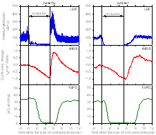
Figure 1: Comparison of the postocclusive reactive
hyperemia in foot after a 5 minute arterial occlusion on thigh
measured by means of three noninvasive methods (NIRS, LDF and
transcutaneous oximetry - TcPO2)
in a healthy volunteer (left) and a diabetic patient with
advanced arterial occlusive disease (right). Hyperemia was by
far more pronounced in healthy foot.
[click on the image to enlarge] |
|
|
|
|
 |
Figure 2: Example of changes in blood flow and
oxygenation in subcutaneous murine tumors after intravenous
injection of antihypertensive hydralazine (left) and after
application of electroporation pulses (right). Perfusion and
oxygenation were reduced in both cases, but the reduction in
perfusion was much faster and more pronounced after
electroporation.
[click on the image to enlarge] |
Bibliography:
- Kragelj R, Jarm T, Miklavcic D. Reproducibility of
parameters of postocclusive reactive hyperemia measured by
near-infrared spectroscopy and transcutaneous oximetry.
Annals Biomed. Eng. 28:168-173, 2000. [PDF
 ] ]
- Kragelj R, Jarm T, Erjavec T, Preseren-Strukelj M, Miklavcic
D. Parameters of postocclusive reactive hyperemia measured by
near-infrared spectroscopy in patients with vascular disease and
in healthy volunteers. Annals Biomed. Eng. 29:311-320,
2001.
[PDF]
- Jarm T, Sersa G, Miklavcic D. Oxygenation and blood flow in
tumors treated with hydralazine: Evaluation with a novel
luminescence-based optic sensor. Techol. Health Care
10: 363-380, 2002. [PDF]
- Jarm T, Cemazar M, Steinberg F, Streffer C, Sersa G, Miklavcic
D. Perturbation of blood flow as a mechanism of anti-tumour
action of direct current electrotherapy. Physiol. Meas.
24: 75-90, 2003. [PDF]
- Jarm T, Kragelj R, Liebert A, Lukasiewitz P, Erjavec T,
Preseren-Strukelj M, Maniewski R, Poredos P, Miklavcic D.
Postocclusive reactive hyperemia in healthy volunteers and
patients with peripheral vascular disease measured by three
noninvasive methods. Adv. Exp. Med. Biol. 530: 661-669,
2003.
|
 |